News
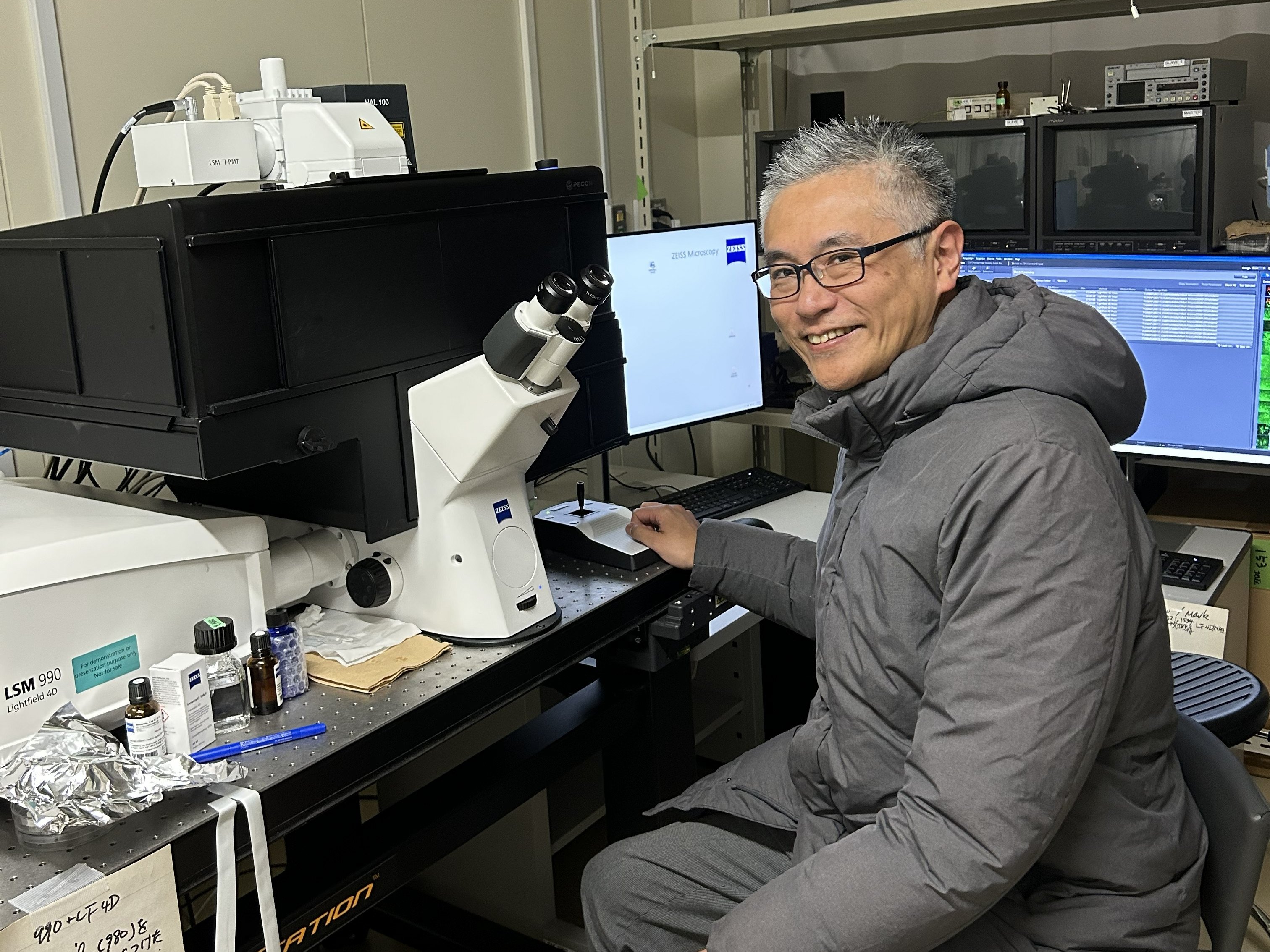
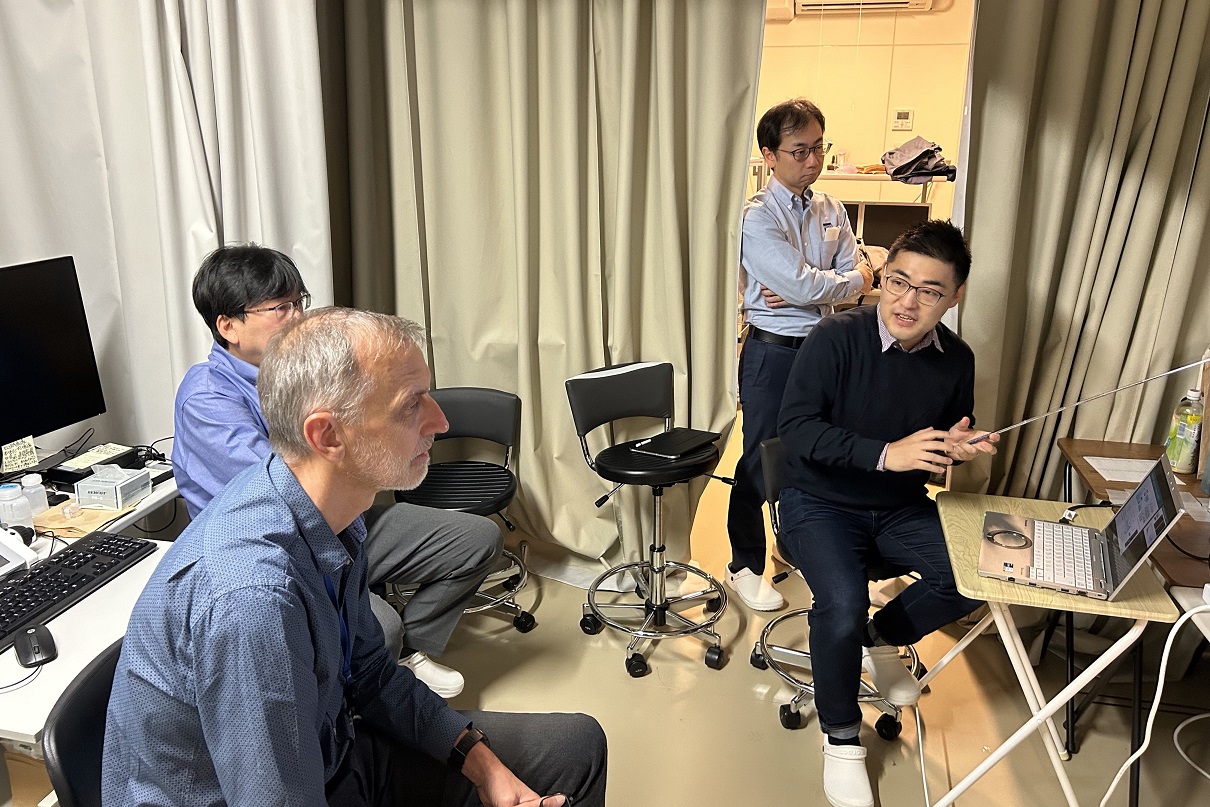
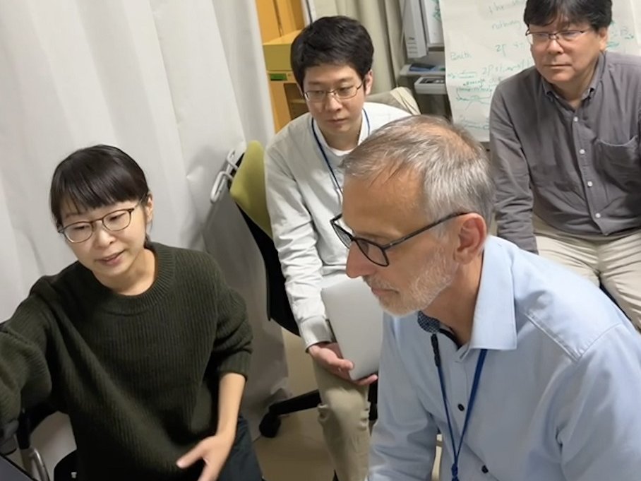
[January 13 - February 5, 2026] LSM990 with Lightfield 4D Demo Sessions
During the demo period, 11 instrument briefing sessions and 10 experiment sessions were conducted. The experiment sessions provided active discussion and valuable feedback.
[February 2, 2026] New publication: Wang et al., J Extracell Biol
New publication by Tanaka group, UTokyo. Lattice light-sheet microscopy showed that tubulovesicular Sonic Hedgehog–Containing Extra-Large Extracellular Vesicles (SHH-XLEVs) with diameters of 600–2000 nm were released from the perinuclear plasma membrane region. Scale bar, 10 μm.
[January 21, 2026] Extension of CRA on ZEISS-iCeMS Innovation Core
ZEISS and iCeMS agreed to extend the activity of ZEISS-iCeMS Innovation Core until October 31, 2026.
[December 12, 2025] New publication: Kumar et al., Plant Nano Biol
New publication by GP group of iCeMS/Graduate School of Engineering, KU. Coriander seed–derived carbon quantum dots were developed as versatile nanomaterials with cross-kingdom functionality, offering potential in both cellular rejuvenation and agricultural nanobiotechnology.
[October 1, 2025] New publication: Farrag et al., JACS
New publication by Uesugi group of iCeMS/Institute for Chemical Research, KU. A novel fluorescent technique was developed to distinguish between liquid and solid states of protein condensates in live cells and isolate distinct populations for omics analysis.
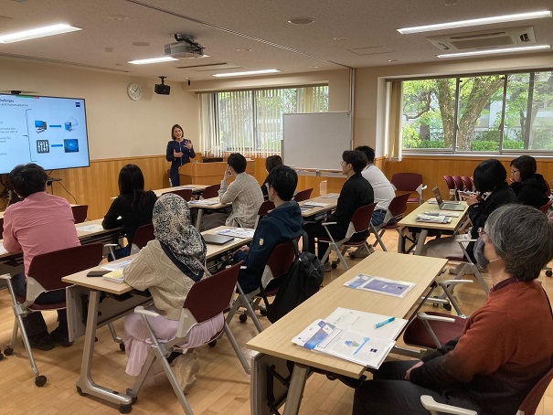
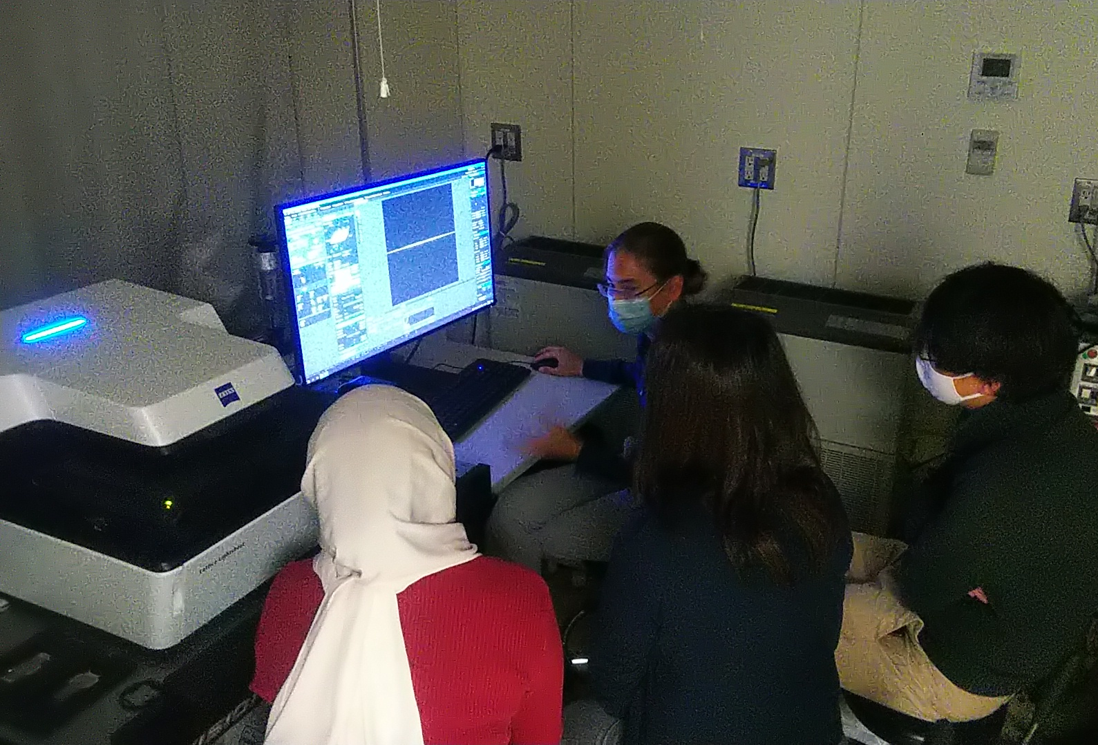
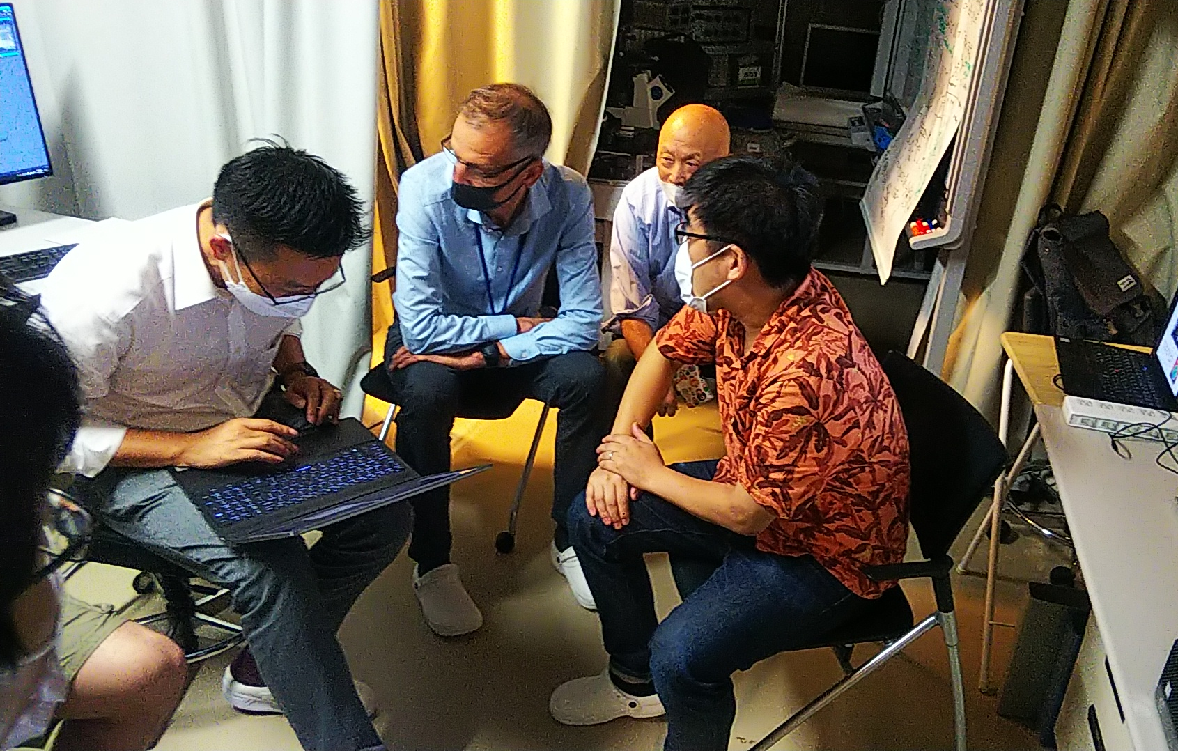
[August 18-20, 2025] 4D Multimodal Imaging Workshop
ZEISS Lightfield 4D acquires large volumes of multicellular specimens in a single snapshot, enabling high-speed tracking of dynamic processes (up to 80 volumes/sec)
or imaging with very low-toxicity over extended periods, whereas Lattice SIM 5 offers different 4D imaging capabilities that can visualize subcellular structures and
track their dynamics at super-resolution.
An online technical seminar on up-to-date 4D imaging technologies and their applications was given by ZEISS on August 18, from 17:00 to 18:00. For demonstration,
short group tours were organized for August 18, and personal sessions for August 19 and 20.
Date & Time
Online Technical Seminar by Carl Zeiss Microscopy via Zoom:
August 18 (Mon), 2025, 17:00-18:00
Dr. Bernhard Zimmermann (moderator: Head of Business Sector Life Sciences)
Dr. Annette Bergter (Lightfield 4D: Solution manager for LSM, Business Sector Life Sciences)
Dr. Xianke Shi (Lattice SIM 5: Senior Manager, Application & Business Development, Life Sciences, Asia Pacific)
Demonstration:
August 18 (Tue) – 20 (Wed)
Group Tour (5 people x 1 hr): August 18
Personal sessions for Lightfield 4D or Lattice SIM 5 with own sample (1-2 hrs): August 19 and 20
Place
Technical seminar:
Onsite Group Zoom Viewing: Rm 119 of Research Bldg. No.1 (#32)
Demonstration:
Rm 305 of Research Bldg. No.1 Annex (#33) and Rm 311 of iCeMS Research Bldg. (#31)
Language
English (seminar); Japanese or English (demonstration)
Flyer
[April 1, 2025] Extension of CRA (Cooperative Research Agreement) on the use of StayGold at the Analysis Center for 2 years
The CRA made effective from Oct. 2, 2023 for 2 years was extended for another 2 years, until Mar. 31, 2027. The iCeMS Analysis Center and Dr. Miyawaki of RIKEN provide the plasmids encoding StayGold fluorescent protein probes and technically support the advanced microscopic observations at the Analysis Center.
[January 21, 2025] New publication: Rodriguez-Reza et al., Curr Biol
New publication by Carlton group of Graduate School of Biostudies, KU. A new model was proposed on how chromosomes measure their own lengths from the crossover sites during meiosis.
[January 16, 2025] New publication: Takato et al., Nat Chem Biol
New publication by Hamachi group of Graduate School of Engineering and iCeMS, KU. Photooxidation-driven proximity labeling for proteome identification (PhoxID) method was developed and applied to neurotransmitter receptor-proximal proteins in the live mouse brain.
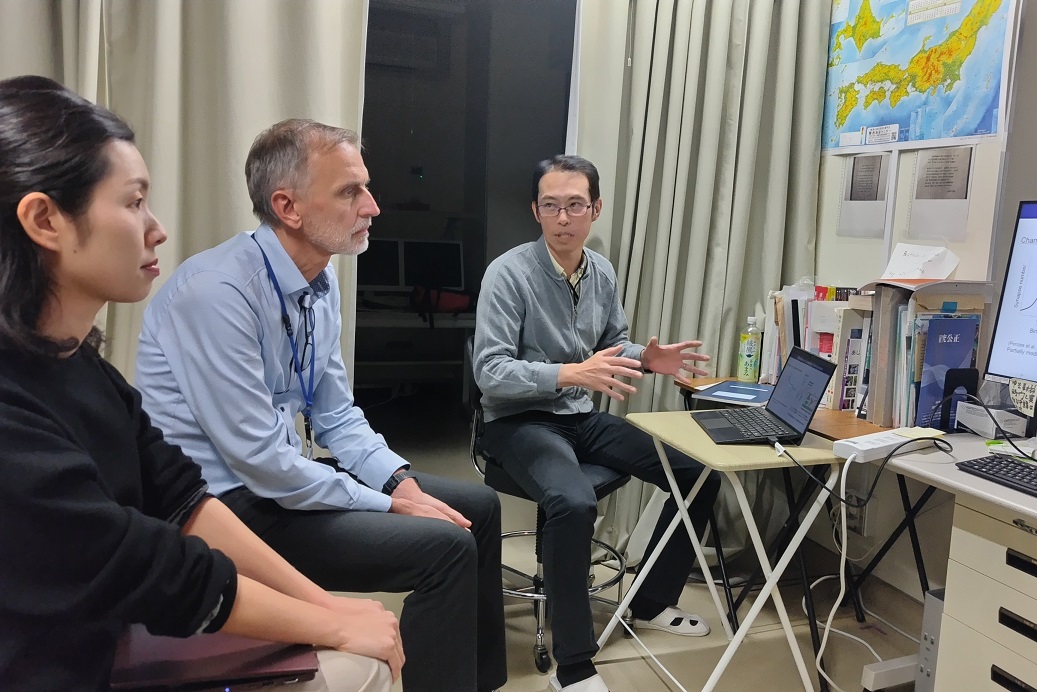
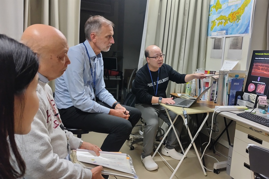
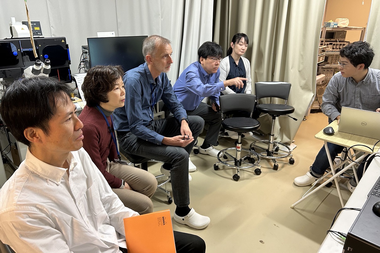
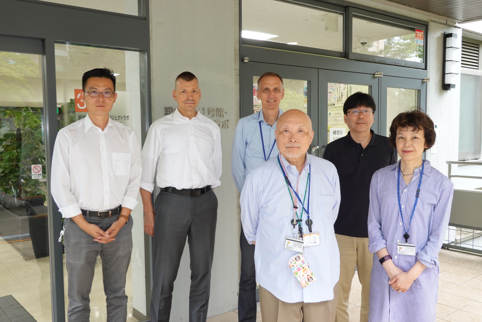
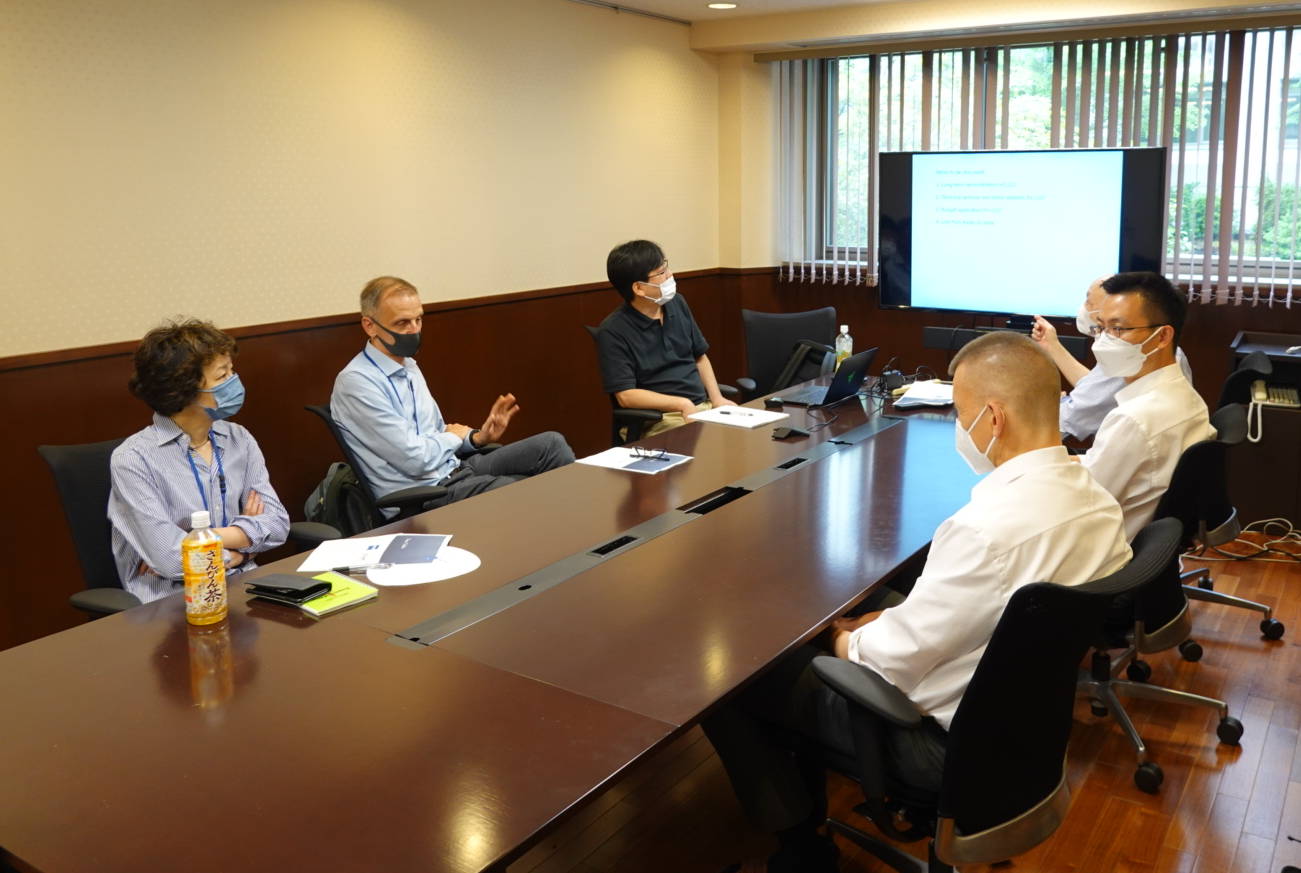
[November 18 and 19, 2024] Meeting and discussion at Kyoto University 2024
ZEISS and iCeMS agreed to extend the activity of ZEISS-iCeMS Innovation Core for 1 year, until October 31, 2025. On this occasion, 11 user group meetings and 2 online meetings to discuss ongoing projects and future collaborations were held at the Innovation Core on November 18th and 19th, 2024.
On 19th afternoon, Dr. Bernhard Zimmermann gave a technical seminar to introduce new hardware and software developments, especially focusing on dynamics measurement features, Dynamic Profiler and Spectral RICS, which were recently made available for LSM980 with Airyscan2.
Schedule
Disucussion with the researchers:
November 18 (Mon) and 19 (Tue)
Meeting by the Innovation Core member:
November 19 (Tue), 13:00-15:00
Technical seminar:
November 19 (Tue), 17:00-18:00
Dr. Bernhard Zimmermann (Head of Business Sector Life Sciences, ZEISS Research Microscopy Solutions, Jena)
[November 15, 2024] New publication: Maki et al., J Cell Sci
New publication by Maki group of Institute for Life and Medical Sciences, KU. Lattice SIM super-resolution microscopy revealed that the nuclear body is encapsulated by a single-stranded (ss)DNA-based molecular complex and the complex upholds the structural integrity of nuclear bodies and their function such as gene transcription and replication.
[August 8, 2024] New publication: Zhou et al., J Cell Biol
New publication by Kengaku group of iCeMS. Nesprin-2 coordinates the dynein complex and kinesin-1 and activates prolonged bidirectional movements of the nucleus along forward-moving microtubules in migrating neurons.
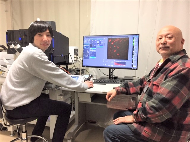
[June 25, 2024] Technical Seminar on FCS and FLIM applications
Dr. Xianke Shi (ZEISS Microscopy Life Sciences, APAC) gave a technical seminar on FCS and FLIM applications provided for ZEISS system, especially focusing on "Dynamic Profiler" software functionality developed for Airyscan detector. It enables detections of molecular concentration, diffusion, and flow dynamics of fluorescent molecules in live cell samples.
[January 24, 2024] New publication: Fukute et al., Commun Biol
New publication by Maki group, Institute for Life and Medical Sciences, KU. Mapping of DNA in underwound state by Lattice SIM^2 revealed that anchoring to nuclear condensates constrains the axial rotation of DNA, facilitating RNAP-driven DNA underwinding in nucleoli.
[December 1, 2023] New publication: Ando et al., Nat Methods
Miyawaki group in RIKEN, in collaboration with the member of ZEISS-iCeMS Innovation Core, developed monomeric StayGold and improved tandem dimer variants. Momomeric StayGold gained higher resistance to strong excitation light and faster maturation than original StayGold. Volumetric imaging of cristae membrane dynamics in a cell-wide manner was performed by Lattice SIM^2 (below: acquisition by Dr. Anna Löschberger and movie processing by Dr. Kirstin Elgass of Carl Zeiss).
[October 2, 2023] CRA (Cooperative Research Agreement) on the use of StayGold, a highly photostable and bright green fluorescent protein, at the Analysis Center
Researchers at KU can be provided with the plasmids containing the sequence of StayGold (and its variants) "free of charge" and "without individual MTA" only when the observations are made with the microscope facility at the Analysis Center. The Analysis Center and Dr. Miyawaki will technically support the research using StayGold probes. The CRA is effective from Oct. 2, 2023 to Mar. 31, 2025.
[August 1, 2023] New publication: Yu et al., Nat Commun
Lattice SIM^2 images were employed for a new publication by Yoshimura Group of Graduate School of Biostudies, KU. The BAR domain-containing protein CIP4 is critical in the actin-dependent asymmetric closing process of clathrin-mediated endocytosis, and the asymmetricity is generated by the self-assembly of the disordered region of CIP4.
[June 7, 2023] New publication: Fujiwara et al., J Cell Biol
Two papers by the Innovation Core member (Fujiwara et al., J Cell Biol), in which Dr. Thomas Kalkbrenner of ZEISS was involved, were added to the publicaion list. An ultrafast camera with single fluorescent-molecule sensitivities was developed and applied to the studies on plasma membrane and focal adhesion structure and function.
[June 2-5, 2023] Lattice Lightsheet 7 Demonstration Sessions
Demonstration sessions for Lattice Lightsheet 7, supported by Mr. Yasuhiko Sato from ZEISS Japan, will take place at ZEISS-iCeMS Innovation Core on June 2-5. Please contact ZEISS-iCeMS Innovation Core for registration.
Date & Time
June 2 (Fri) - 5 (Mon), 1 - 2 sessions per day, including long-term observations
Place
ZEISS-iCeMS Innovation Core, Room 305 of Research Bldg. No.1 Annex (#33)
[April 18 and 19, 2023] Demonstration Seminar on arivis Pro
A demonstration seminar (onsite-online hybrid) on arivis Pro (formerly vision4D), an image analysis software suitable for volumetric 3D/4D data sets acquired with lightsheet and super-resolution microscopes was held on April 18 (Tue), 10:00-11:00. Dr. Delisa Garcia (ZEISS arivis, UK) presented new features of arivis Pro, such as efficient analysis workflow of 3D/4D data sets, neuron tracing, and AI-based image processing.
On April 18 afternoon and April 19, technical support and discussion sessions for reserchers from iCeMS and other departments of Kyoto University were arranged.
[February 6-8, 2023] Lattice Lightsheet 7 Demonstration Sessions
Demonstration sessions for Lattice Lightsheet 7, supported by Mr. Yasuhiko Sato from ZEISS Japan, will take place at ZEISS-iCeMS Innovation Core on February 6-8. Please contact ZEISS-iCeMS Innovation Core for registration.
Date & Time
February 6 (Mon) - 8 (Wed), 1 - 2 sessions per day, 3 hrs each
Place
ZEISS-iCeMS Innovation Core, Room 305 of Research Bldg. No.1 Annex (#33)
[January 19-20, 2023] Lattice Lightsheet 7 Demonstration Sessions
Demonstration sessions for Lattice Lightsheet 7, supported by Mr. Yasuhiko Sato from ZEISS Japan, will take place at ZEISS-iCeMS Innovation Core on January 19 and 20. Please contact ZEISS-iCeMS Innovation Core for registration.
Date & Time
January 19 (Thu) and 20 (Fri), 2 sessions per day, 3 hrs each
Place
ZEISS-iCeMS Innovation Core, Room 305 of Research Bldg. No.1 Annex (#33)
[January 5, 2023] New publication: Ogasawara and Ueda, J Chem Biol
New publication by iCeMS member, Ogasawara and Ueda, J Chem Biol, was added to the publicaion list. They revealed important roles of two proteins (ABCA1 and Aster-A) in maintaining an appropriate distribution of cholesterol inside cells by high-resolution imaging, which provided new insights into how cells achieve cholesterol homeostasis.
[December 13-16, 2022] Lattice Lightsheet 7 Workshop
A workshop for Lattice Lightsheet 7 took place at ZEISS-iCeMS Innovation Core on December 13-16.
Lattice Lightsheet 7 provides lattice lightsheet fluorescence microscopy for live cell imaging with minimum photodamage
while allowing your use of standard glass bottom dishes/well plates. With the automated system calibration and easy-to-use interface,
volumetric imaging of cellular events ranging from dynamics of subcellular structures to multicellular interactions
over hours and days becomes available to everyone.
Dr. Kirstin Elgass (Business Sector Life Sciences, ZEISS Research Microscopy Solutions, Jena, Germany) gave an online seminar on
Lattice Lightsheet technology and its applications to various live-cell imaging on December 13, 16:00-17:00.
On the next three days, onsite demonstration sessions using own sample (about 2 hrs each) were arranged.
Date & Time
Technical seminar:
December 13 (Tue), 2022, 16:00-17:00
Demonstration:
December 14 (Wed) – 16 (Fri), 3 sessions per day, 2 hrs each
Place
Technical seminar: Online via Zoom
Demonstration: ZEISS-iCeMS Innovation Core, Room 305 of Research Bldg. No.1 Annex (#33)
Language
English (seminar); Japanese or English (demonstration)
Flyer
You will have access to the system installed at ZEISS-iCeMS Innovation Core for several months. Please contact
ZEISS-iCeMS Innovation Core if you get interested in long-term use of the system.
[October 31-November 4, 2022] "Applicathon" Workshop at Kyoto University
The 1st "Applicathon" Workshop to comprehensively evaluate the performance of 4 ZEISS microscopes available at the Innovation Core (LSM 880, LSM 980, Elyra 7, and Lattice Lightsheet 7) was organized. ZEISS engineers and operators from Germany, Singapore, Korea, and Japan and 7 research groups from iCeMS and other departments of Kyoto University worked together to observe various kinds of samples on the different systems and evaluate which system is most suitable for each sample.
[October 31, 2022] Lattice Lightsheet 7 is under demonstration for evaluation.
Lattice Lightsheet 7 was installed for long-term demonstration and evaluation. Please see the specification from the
Equipment menu.
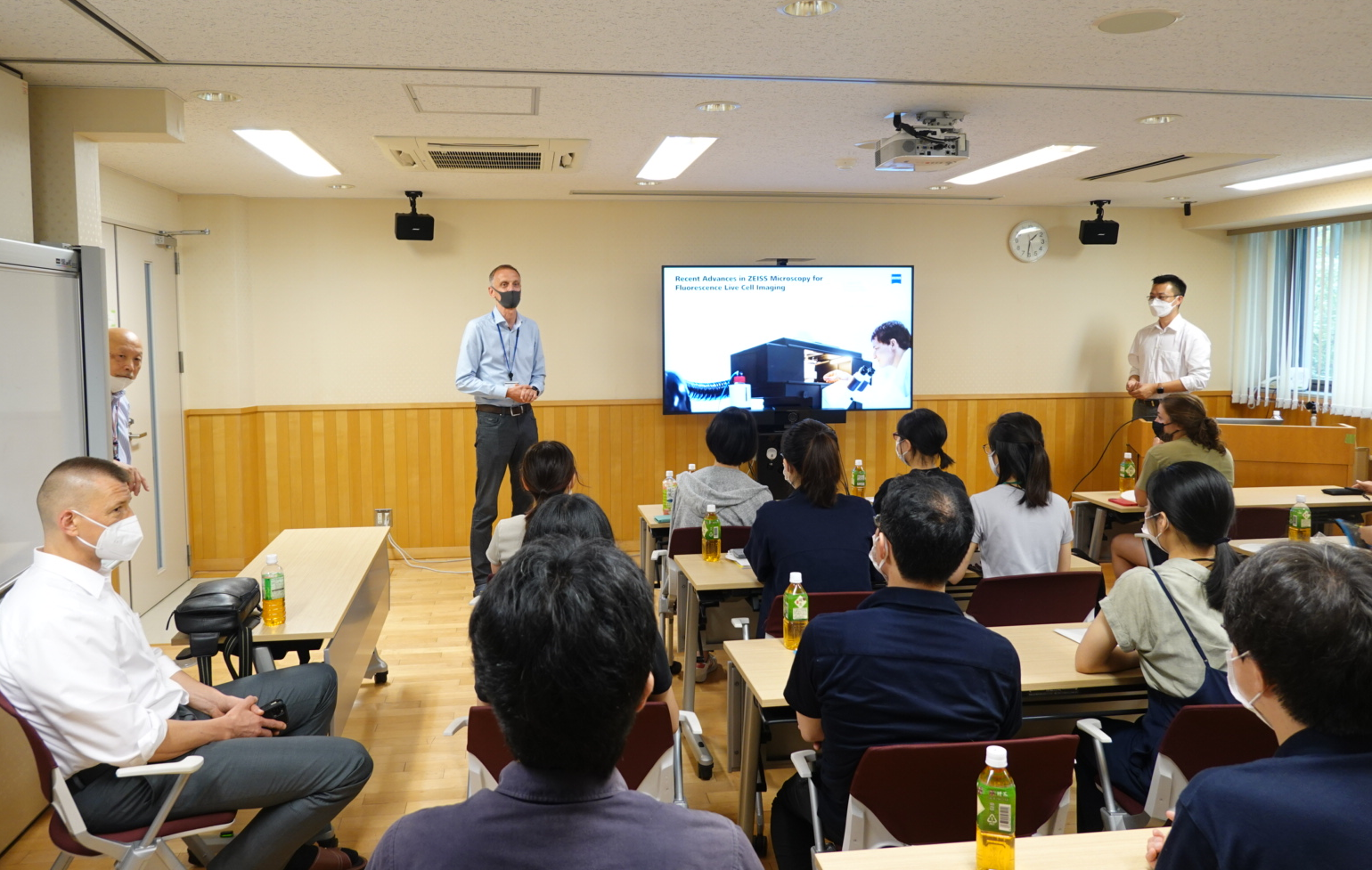
[July 4 and 5, 2022] Meeting and discussion at Kyoto University 2022
ZEISS and iCeMS-KUIAS agreed to extend ZEISS-iCeMS Innovation Core based on the collaborative research agreement by two years, until October 31, 2024.
On this occasion, a meeting to discuss about the future collaboration was held at the iCeMS on July 4 and 5, 2022.
Dr. Xianke Shi gave a technical seminar entitled "Recent Advances in ZEISS Microscopy for Fluorescence Live Cell Imaging".
In addition, 11 research groups from iCeMS and other departments of Kyoto University presented their research and discussed current problems and future technical requirements.
Schedule
Meeting by the Innovation Core member:
July 4 (Mon), 2022, 11:00-13:00
Technical seminar:
July 4 (Mon), 2022, 13:30-14:15
Dr. Xianke Shi (ZEISS Microscopy Life Sciences, APAC)
Recent Advances in ZEISS Microscopy for Fluorescence Live Cell Imaging
Disucussion with the researchers:
July 4 (Mon) and July 5 (Tue)
[April 26, 2022] New publication on StayGold, super-stable fluorescent protein: Hirano et al., Nat Biotechnol
New publication by Miyawaki group (RIKEN) was added to the publicaion list. Images by Elyra 7 were used.
[March 28, 2022] Online Seminar (via Zoom): Towards fast and gentle super-resolution microscopy
Super-resolution fluorescence microscopy is rapidly evolving the field of biomedical research. Recent technical developments are mainly focused on fast and gentle observation of dynamic cellular structures ranging from mesoscale (20~500 nm) molecular assemblies to highly organized cell organelles.
In this online seminar, first, the scientific lecture on the development of novel fluorescent proteins to improve observations of various cellular processes is given by Dr. Atsushi Miyawaki (RIKEN CBS). Next, the technical lecture on the features of two ZEISS super-resolution microscopes “Elyra 7 with Lattice SIM2” and “LSM 980 with Airyscan 2”, available at ZEISS-iCeMS Innovation Core, is given by Dr. Klaus Weisshart (ZEISS Jena, Germany).
Elyra 7 was just installed as shared equipment of MaCBES (Main Campus Base of Equipment Support), Kyoto University in December 2021. After the seminar, we will have onsite demonstration sessions for Elyra 7 using your samples (about 2 hrs each) on March 29, 30, and 31.
Please register for the online seminar and the onsite demonstration session by March 18 from the Registration URL below. Zoom meeting information will be announced by e-mail.
Date: March 28 (Mon), 2022
14:00-14:45
Atsushi Miyawaki (Riken CBS)
Continuity in Space and Time
14:45-15:45
Klaus Weisshart (ZEISS Jena, Germany)
Latest developments in confocal and widefield based super-resolution microscopy using the example of Carl Zeiss Airyscan 2 and ELYRA 7
Language: English
Registration:
https://forms.gle/Xtr4skoksw4zSpK67 (closed)
Flyer
[December 2, 2021] Elyra 7 with Lattice SIM^2 was installed.
Elyra 7 with Lattice SIM^2 was installed as shared eqipment of
MaCBES (Main Campus Base of Equipment Support). Please see the specification from the
Equipment menu. We will have a technical seminar on Lattice SIM^2 and demo sessions in next January.
[September 28, 2021] New publication: Minakawa et al., J Extracell Vesicles
New publication by Innovation Core user, Minakawa et al., J Extracell Vesicles, was added to the publicaion list.
[June 1, 2021] Publication list updated.
Publication list was updated, and the links to the Citations (by Dimensions) and Altmetric were added.
[April 7, 2020] iCeMS Specially Appointed Professor Fumiyoshi Ishidate, member of
ZEISS-iCeMS Innovation Core, was selected to receive the "Outstanding Support for Research Award"
of the "Commendation for Science and Technology by the Minister of Education, Culture, Sports, Science and Technology"
for 2020.
The "Commendation for Science and Technology by the Minister of Education, Culture, Sports, Science and Technology
(
科学技術分野の文部科学大臣表彰)" honors those who have made remarkable achievements in the field of science and technology,
and the "Outstanding Support for Research Award (
研究支援賞)" is a newly established award given to those who supported
the progress of research and development activities though their highly professional knowledge and skills. Prof. Ishidate
is one of the first winners of this honorable award.
Prof. Ishidate has made great contributions to iCeMS imaging facility for these 10 years, especially as a mentor of
live cell imaging using ZEISS microscopes. He provided one-on-one training sessions to a cumulative total of 600
researchers inside and outside the university. In addition, through the collaboration with iCeMS Kageyama group, he
developed the "long-term microscopic observation technique for weak signals in living cells" taking advantage
of his deep knowledge of live cell samples and skills to operate advanced confocal microscopes. Using the developed
technique, time-lapsed imaging of weak signals from the fate-determination transcription factors in neural progenitor
cells was successfully performed for the first time. It resulted in the finding that the multipotent state correlates
with oscillatory expression of several fate-determination factors, whereas the differentiated state correlates
with sustained expression of a single factor (Imayoshi et al., 2013, 2015), which attracted worldwide attention.
Prof. Ishidate also made continued efforts to establish the long-standing partnership between iCeMS and ZEISS German
headquarters, and based on the partnership, we were able to launch the ZEISS-iCeMS Innovation Core in 2019.
Congratulations, Ishidate-san!
References
1. Imayoshi I, Isomura A, Harima Y, Kawaguchi K, Kori H, Miyachi H, Fujiwara T, Ishidate F, Kageyama R. Oscillatory
control of factors determining multipotency and fate in mouse neural progenitors. Science 342: 1203-1208 (2013).
2. Imayoshi I, Ishidate F, Kageyama R. Real-time imaging of bHLH transcription factors reveals their dynamic control
in the multipotency and fate choice of neural stem cells. Front Cell Neurosci 9: 288 (2015).
[January 27-29, 2020] ZEISS Elyra 7 with Lattice SIM Workshop
A workshop for Elyra 7 with Lattice SIM, newly developed lattice structured illumination-based
super-resolution microscope system, took place at ZEISS-iCeMS Innovation Core on January 27-29.
In Lattice SIM, the sample area is illuminated with a lattice pattern instead of grid lines. The lattice
pattern gives higher contrast and 2x higher sampling efficiency compared with conventional SIM, so that you
need less illumination. This is a breakthrough in light efficiency, enabling fast live imaging of 3D volumes
at excellent resolutions (120 nm in XY and 300 nm in Z) while minimizing photodamage.
Dr. Bernhard Zimmermann (Senior Director Business Sector Life Sciences, ZEISS Research Microscopy Solutions,
Jena, Germany) gave a seminar on Lattice SIM technology and its comparison to other super-resolution
methods. After the seminar, demonstration sessions using own samples (about 2 hrs each) were arranged.
Date & Time
Technical seminar:
January 27 (Mon), 2020, 13:30-14:30
Demonstration:
January 27 (Mon), 2020, 1 session after the seminar
January 28 (Tue), 2020, 3 sessions
January 29 (Wed), 2020, 3 sessions
Place
Technical seminar: Room 119 of Research Bldg. No.1 (#32) at Main Campus of Kyoto University
Demonstration: ZEISS-iCeMS Innovation Core, Room 305 of Research Bldg. No.1 Annex (#33)
Language
English
Flyer
You will have access to the system installed at ZEISS-iCeMS Innovation Core for several months. Please contact
ZEISS-iCeMS Innovation Core if you get interested in long-term use of the system.
[December 23 and 24, 2019] ZEISS LSM 980 with Airyscan2 Briefing Session
A briefing session for ZEISS LSM 980 with Airyscan2, recently installed as a common facility in iCeMS Analysis
Center/ZEISS-iCeMS Innovation Core, was held on December 23 and 24.
The microscope provides the capability of fast and gentle 4D super-resolution imaging over large view fields.
The difference between Airyscan2 (confocal-based super-resolution) and ZEISS Elyra 7 with Lattice SIM (newly
developed lattice structured illumination-based super-resolution suitable for live imaging) was also
discussed.
Masako Yamaguchi (ZEISS Research Microscopy Solutions) gave technical seminar and quick demonstration at the
microscope system.
Date & Time
Slot 1: Monday, December 23, 2019 13:00-14:00 (Japanese)
Slot 2: Monday, December 23, 2019 15:00-16:00 (Japanese)
Slot 3: Tuesday, December 24, 2019 13:00-14:00 (Japanese)
Slot 4: Tuesday, December 24, 2019 15:00-16:00 (English)
Place
Technical seminar: Room 119 of Research Bldg. No.1 (#32) at Main Campus of Kyoto University
Demonstration: ZEISS-iCeMS Innovation Core, Room 304 of Research Bldg. No.1 Annex (#33)
Flyer
[October 28, 2019] ZEISS-iCeMS Innovation Core Founding Commemorative
Symposium & Opening Ceremony
ZEISS-iCeMS Innovation Core Founding Commemorative Symposium was hosted by iCeMS on October 28.
Ko-ichiro Tanaka
(Kyoto Univ Dept of Physics / iCeMS)
Terahertz Imaging and Nearfield Microscopy
Naoki Watanabe
(Kyoto Univ Dept of Medicine)
Single-Molecule Dissection of Physico-Biochemical Coupling in Actin System
Ryoichiro Kageyama
(Kyoto Univ Inst for Frontier Life & Med Sci / iCeMS)
Live Imaging of Dynamic Expression of the Mouse Segmentation Clock Gene Hes7
Date & Time
October 28, 2019
13:00-15:00
Venue
KUIAS/iCeMS Main Building 2F Seminar Room (A207)
Flyer
After the Symposium, Opening Ceremony of ZEISS-iCeMS Innovation Core and Signing of the Collaborative Research
Agreement between ZEISS and KUIAS/iCeMS Kyoto University were holded.
Link to Press Release by ZEISS Research Microscopy Solutions
Link to Kyoto University News
Link to iCeMS News (Japanese)